ANT 326 Human Osteology
Tuesday, September 11, 2012
Finishing up on Appendicular Skeleton I: Humerus, Radius, Ulna
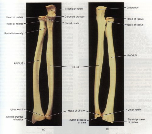
Radius: Here is a description of the 8 features listed on your
handout/notes from class. The image below has some of these features
labeled. Please refer to Bass and White et al. and come to the lab to see
the bones and familiarize yourself with the location of these features.
- Head: Located proximally, it's the round portion that
rotates along the capitulum of the humerus so that the hand can turn from
palm-up to palm-down.
- Neck: The tapered portion inferior to the head, yet still
on the proximal portion of the bone.
- Radial tuberoisty: The bump inferior to the neck.
It's for muscle attachment.
- Nutrient foramen: Hole or opening for a major blood vessel.
Its placement varies along the diaphysis of the bone.
- Interosseous crest: The sharp ridge of bone running along
the diaphysis ("inter" = between, "osseous" = bone; it's the sharp region
where muscles attach between the radius and the ulna). It's not
pictured below, but you can see it in Bass and White et al.
- Styloid process: This is a tiny point, distally.
Think "stylus" as in "pen" and a pen-point. More than one bone has a
styloid process (e.g., ulna, pictured below).
- Dorsal tubercles: Little bumps on the dorsal (back) side of
the radius, distally. Please see Bass and White et al.
- Ulnar notch: This is an indentation on the lateral side of the
distal portion of the radius; it is named the "ulnar" notch because it is a
facet for articulation with the head of the ulna.
- Oftentimes, features involved in articulations (bone connections) have
names that relate to what is articulating with them. Thus, the radius
has a feature--the ulnar notch--so named because that's where the head of
the ulna articulates.
- Features that are for muscle attachment (not connections with other
bones) may have names that refer to the bone itself, such as the radial
tuberosity on the radius, which is bump for muscle attachment.

Image from:
http://www.cas.muohio.edu/~meicenrd/mudescd/Bot630DProjects/2003Botany630WProjects/Bones/RADIUS&ULNA.JPG
- Please note the plural for radius is radii (ray-dee-eye); and the plural
for humerus is humeri (hyoo-mur-eye).
Ulna: Here I will cover the 8 features listed on your handout;
you can refer to the photo above and please also use your White textbook and
Bass field guide. Remember to come to the lab to practice "siding" bones
and locating features ("siding" = determining if a paired bone is a left or
right). The plural of ulna is ulnae (ul-nye in proper Latin pronounciation;
but, you'll often hear it pronounced "uhl-nee").
- Olecranon: This is the feature superior on the proximal
portion of the bone; it's the part of the bone that makes that bump we call
the elbow.
- Semilunar notch: This is the half-moon ("semi" means half
and "lunar" means moon) shaped indentation; it's where the ulna articulates
with the trochlea of the humerus. Note that in the
photograph above it's called the "trochlear notch". We are learning
the "semilunar notch" variation. Recall that many terms are
synonymous; however, to keep things simpler, please use the preferred term
from class.
- Coronoid process: This is a sharp projection of bone
anteriorly located on the proximal portion of the bone. Please pay careful
attention to the "n" in "coronoid" and link it to the "n" in "ulna" because
soon we'll learn other features of other bones that have very similar
sounding names (i.e., coracoid process, conoid process, etc.).
- Radial notch: This is the indentation just lateral to the
coronoid process on the proximal portion of the bone. Similar to the
ulnar notch of the radius, the radial notch of the ulna is named for the
bone/feature that articulates with it. Thus, the radial notch is for the
head of the ulna. Note in the photograph above that the head of the
ulna is distal. We must think of "heads" of bones as features
named for the shape: roundish, as opposed to location (i.e.,
the head is not always superior or proximal or at the top; it's a "head"
because it's "head" or "round" shaped).
- Nutrient foramen: Similar to the radius, this is the
opening for the blood vessel and it's oriented such that it's toward the
elbow. Therefore, the opening in the bone where the blood vessel
enters is inferior (it's also inferior on the radius; but recall from class
that it's superior in the humerus). Also, please note that the
nutrient foramen is not seen/labeled in the above photograph. You have
to be fairly close to the bone to see it well as it's tiny.
- Interosseous crest: Similar to the feature of the same name
on the radius, this is the long sharp ridge of bone traveling along the
lateral side of the diaphysis (shaft). Muscles involved in pronation
and supination (turning the hand palmar and dorsal) attach along these
crests of the ulna and radius. This featuer is not labeled in the
above photograph; however, your White text and Bass field guide will show
it.
- Styloid process: Located on the head of the ulna (distal),
this is the projecting point lateral near your wrist. Remember, just like
the styloid process of the radius, the styloid process of the ulna is like a
pen-tip (think of "le stylo" or pen in French).
- Head: Round part of the bone. The head of the ulna is
unusual in that it is distally located.